![]()
A Guide to Brain Anatomy
Call me at 800-992-9447
You are now on waiting.com, the Coma Waiting Page, a webpage that I co-wrote in 1997 to provide immediate assistance to the families of someone waiting to wake up from a brain injury coma. When I started the waiting.com project, there was no information about coma or severe brain injuries on the internet. I named it waiting.com because it was written to be read in an emergency room, to provide hope and a connection to others who had also waited for someone to awake from a coma, resulting from a traumatic brain injury.
I’m not a doctor. I’m a lawyer. I make my living representing those who have suffered brain injuries because of the wrongful conduct of others. I had been writing about TBI for publication for several years before I discovered what the internet could mean for education. I was editor of a couple of TBI newsletters, including the Brain Injury Association of Wisconsin’s newsletter. When I realized that I could create a page that could be available to almost anyone, I realized that I had found my calling.
Since 1996, I have written thousands of pages of internet content about traumatic brain injury. The parts of that content that relate to brain anatomy and function are on the next few pages. As you click through, there will be links to many more pages on specific issues that you or your loved ones will need help with. I hope we have laid out this information is such a way as you can get access to the specific help that you need.
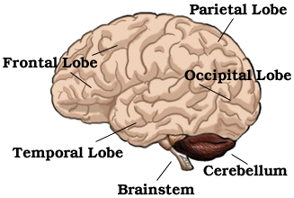
Brainstem - The lower extension of the brain where it connects to the spinal cord. Neurological functions located in the brainstem include those necessary for survival (breathing, digestion, heart rate, blood pressure) and for arousal (being awake and alert).
Most of the cranial nerves come from the brainstem. The brainstem is the pathway for all fiber tracts passing up and down from peripheral nerves and spinal cord to the highest parts of the brain. Click Here To Return To Diagram
Cerebellum - The portion of the brain (located at the back) which helps coordinate movement (balance and muscle coordination). Damage may result in ataxia which is a problem of muscle coordination. This can interfere with a person's ability to walk, talk, eat, and to perform other self care tasks. Click Here To Return To Diagram
Frontal Lobe - Front part of the brain; involved in planning, organizing, problem solving, selective attention, personality and a variety of "higher cognitive functions" including behavior and emotions.
The anterior (front) portion of the frontal lobe is called the prefrontal cortex. It is very important for the "higher cognitive functions" and the determination of the personality.
The posterior (back) of the frontal lobe consists of the premotor and motor areas. Nerve cells that produce movement are located in the motor areas. The premotor areas serve to modify movements.
The frontal lobe is divided from the parietal lobe by the central culcus. Click Here To Return To Diagram
Occipital Lobe - Region in the back of the brain which processes visual information. Not only is the occipital lobe mainly responsible for visual reception, it also contains association areas that help in the visual recognition of shapes and colors. Damage to this lobe can cause visual deficits. Click Here To Return To Diagram
Parietal Lobe - One of the two parietal lobes of the brain located behind the frontal lobe at the top of the brain.
Parietal Lobe, Right - Damage to this area can cause visuo-spatial deficits (e.g., the patient may have difficulty finding their way around new, or even familiar, places).
Parietal Lobe, Left - Damage to this area may disrupt a patient's ability to understand spoken and/or written language.
The parietal lobes contain the primary sensory cortex which controls sensation (touch, pressure). Behind the primary sensory cortex is a large association area that controls fine sensation (judgment of texture, weight, size, shape).Click Here To Return To Diagram
Temporal Lobe - There are two temporal lobes, one on each side of the brain located at about the level of the ears. These lobes allow a person to tell one smell from another and one sound from another. They also help in sorting new information and are believed to be responsible for short-term memory.
Right Lobe - Mainly involved in visual memory (i.e., memory for pictures and faces).
Left Lobe - Mainly involved in verbal memory (i.e., memory for words and names). Click Here To Return To Diagram
 |
|
|
|
Attorney Gordon S. Johnson, Jr.
![]()
E-mail to: waiting.com
For legal questions call toll free:
1-800-992-9447
copyright ©2002 - 2013 Attorney Gordon S. Johnson, Jr., All rights reserved. For more on Attorney Gordon Johnson
http://gordonjohnson.com